The de Winter Electrocardiogram Pattern Evolving From Hyperacute T Waves. This is a de Winters T-wave and diagnostic of near occlusion of the LAD.
De Winter T Wave Litfl Ecg Library Diagnosis
Represents approximately 2 of LAD occlusions.
. The classic teaching is ST-segment elevation myocardial infarction STEMI is defined as symptoms consistent with acute coronary syndrome ACS new ST-segment elevation at the. The maximal amplitude of the positive tall T wave was 9 3 mm. This specific ECG pattern is seen in relatively young predominantly male and those with higher incidence of.
However it is often unrecognized by physicians. De Winters T Waves - REBEL EM - Emergency Medicine Blog. Optimizing electrocardiogram interpretation and clinical decision-making for acute ST-elevation myocardial infarction.
These patients are suffering occlusion myocardial infarction OMI and require immediate reperfusion therapy. The maximal amplitude of the STD was 3 2 mm. This pattern should be treated as being equivalent to an anterior STEMI.
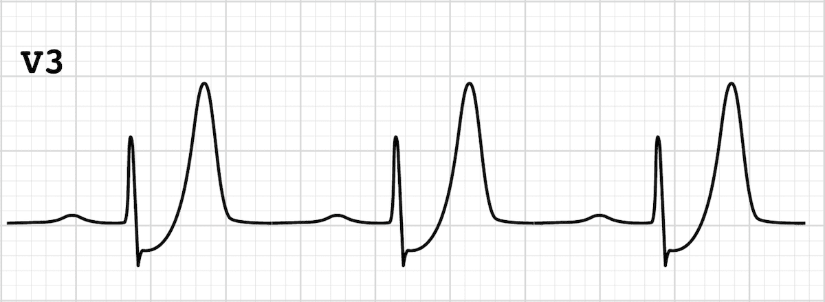
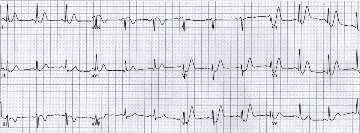
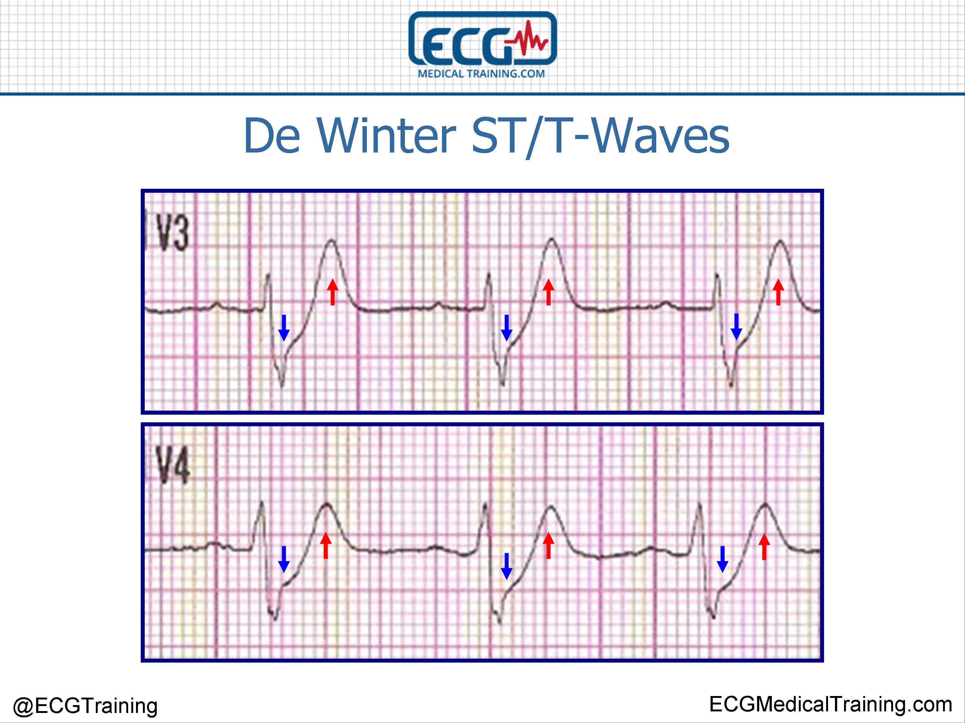
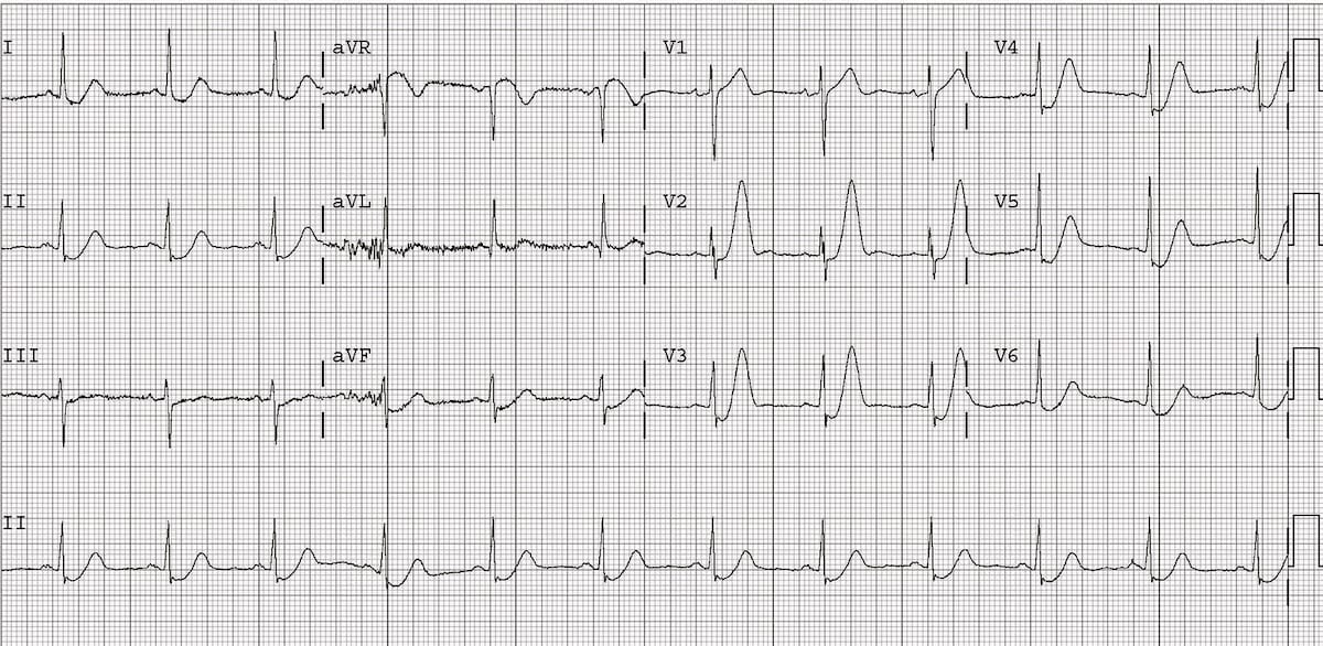
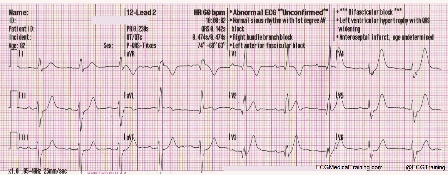
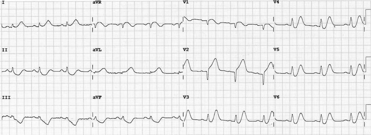
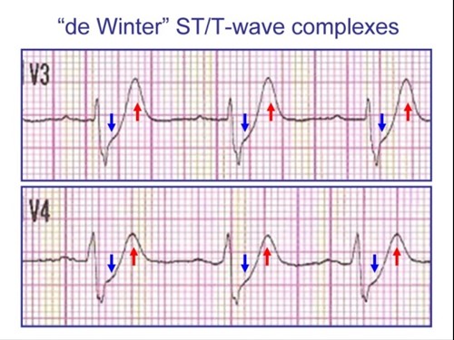
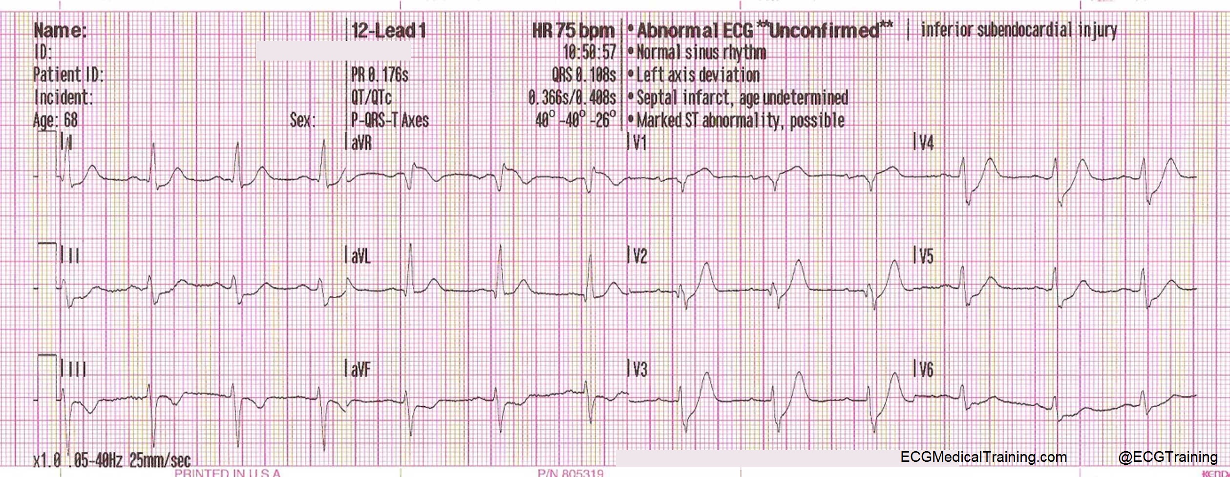
The de Winter electrocardiographic ECG pattern was characterized by upsloping STsegment depressions tall and positive symmetrical T waves in precordial leads. This ECG pattern is commonly referred to as Wellens sign. Tall often very prominent T waves in the precordial leads.
Approximately 10 of patients with acute coronary syndromes have Wellens syndrome. First reported by Dutch Professor of Cardiology Robbert J. Changes are dynamic as you would expect with ACS see Example 3 below Suspicious for proximal occlusion of the LAD.
The electrocardiogram ECG is one of the most useful diagnostic studies for identification of acute coronary syndrome ACS and acute myocardial infarction AMI. T waves associated with De Winters are often referred to as being. Earlier percutaneous coronary intervention PCI.
The heart rate when recording the ECG with de Winter pattern was 74 18 bpm. V3 also has ST depression with a hyperacute T-wave very suggestive of de Winters T-waves. A new ECG sign of proximal LAD occlusion.
Characterized by 1-3 mm of ST-depression with upright symmetrical T-waves. The de Winter electrocardiogram pattern is a transient electrocardiographic phenomenon that presents at early stage of ST-segment elevation myocardial infarction Clin Cardiol 41 2018 pp. Besides the morphology of upsloping or nonupsloping ST depression STD may have different significance of severity and prognostication.
Very slight 05-1mm ST elevation in lead aVR. The de Winter ECG pattern was first described in 2008 as an ST elevation equivalent 3 This pattern was shown as a 1-3 mm upsloping ST-segment depression at V 1-V 6 that continued into tall positive symmetrical T waves. Latest addition to the world of ECG after Bruadas and Wellens.
De Winter in 2008 the de Winter ECG pattern is an anterior STEMI equivalent that presents without obvious ST segment elevation. 75 of these will develop massive anterior myocardial infarction with a high risk of developing heart failure unless revascularization is carried out expeditiously. Most patients showed a J point rise of 1-2 mm at lead aVR mild depression at inferior wall ST segments and normal or slightly elongated.
Rokos I et al. More than 1mm of ST depression in the precordial leads. De Winter R et al.
De Winter Rd et al A new sign of proximal lad occlusion NEJM 2008359. Appropriate cardiac cath lab activation. Onset to recording the de Winter pattern was 99 79 min.
The de Winter ECG pattern is a recently-described STEMI equivalent that emergency physicians and paramedics must be aware of. The De Winter ECG pattern has been reported to indicate acute left anterior descending coronary artery occlusion and is often considered to be an ST elevation myocardial infarction STEMI equivalent. De Winter STT-Waves.
ECG characteristics of De Winters T waves include. Now there is no ST elevation in V2 but there is a hyperacute T-wave. It reminded me that many believe due to the assertions in the original de Winters article that de Winters waves are stable.
Table 1 shows the ECG and angiographic findings of the two groups. The distinct ECG pattern was identified in approximately 2 of patients with LAD stenosis in that study also. In 2008 as a new electrocardiographic EKG pattern of acute proximal left anterior descending coronary artery LAD occlusion.
In 2008 de Winter et al described an ECG pattern suggesting that it should be considered an ST-elevation myocardial infarction STEMI equivalent de Winter Verouden Wellens Wilde 2008 with the potential to predict critical stenosis or occlusion of the left anterior descending coronary artery LAD. Timely recognition of this pattern is crucial. Up to 10 cash back The de Winter ECG pattern characterized by 13-mm upsloping ST-segment depression at the J point in leads V1 to V6 that continues to tall positive symmetrical T waves is identified in only 2 of the patients with acute MI.
These patients were more likely to be younger and male and tended to have hypercholesterolaemia. We aimed to investigate the morphology of the De Winter ECG pattern and evaluate the test characteristics of the De Winter pattern for the diagnosis of acute coronary occlusion. De Winter electrocardiograph ECG pattern signifies proximal left anterior descending coronary artery LAD occlusion and extensive anterior myocardial infarction and it is found in about 2 of patients with proximal LAD occlusion.
ECG abnormality described by de Winter et al. The ST depression persists. The reported positive predictive value PPV for the de Winter ECG pattern to predict an acute left anterior descending artery LAD lesion is inconsistent.
De Winters sign was first described by de Winter et al. These patients typically have critical stenosis of the LAD requiring emergent PCI or thrombolysis. The de Winter pattern could be confused with hyperacute T-waves which occur within minutes of coronary artery occlusion and progress rapidly to classical ST elevation myocardial infarction STEMI pattern.
In fact the title was Persistent precordial hyperacute T-waves signify proximal left anterior descending artery occlusion The authors based this idea of persistence. An electrocardiographic finding suggestive of impending myocardial infarction the de Winters pattern or de Winters T-waves describes an abnormality thought to be indicative of acute occlusion of the proximal left anterior descending coronary artery LAD 2.
De Winter St T Waves Ecg Medical Training
De Winter T Wave Litfl Ecg Library Diagnosis
De Winter St T Waves Ecg Medical Training

De Winter T Wave Litfl Ecg Library Diagnosis
De Winter T Wave Litfl Ecg Library Diagnosis
Dewinter S T Waves
De Winter T Wave Litfl Ecg Library Diagnosis
De Winter St T Waves Ecg Medical Training
0 comments
Post a Comment